Phantoms
ABGT provides a complete range of phantoms and training models for medical imaging, including ultrasound, PET/SPECT, radiotherapy, and angiography. With realistic phantoms for training and quality control, these products ensure accuracy and reliability, essential for optimizing clinical workflows and education.
LUNGMAN – Chest Phantom for Radiology and Tomography
The LUNGMAN phantom is a full-size, accurate, and high-quality anatomical model designed for conventional radiology and computed tomography. It precisely reproduces the thickness and composition of the human thorax, with synthetic tissues and bones that faithfully simulate X-ray absorption.
- Radiography: Enables realistic 3D images from any projection, with accurate contrast of bones and vessels.
- Computed Tomography: Proper positioning for image acquisition, allowing spatial identification of pulmonary vessels and evaluation of automatic detection systems.
- Chest Plate: Increase thorax thickness to simulate different body volumes, adjusting radiation absorption.
Components for Radioisotope: Compatible with CT and PET image fusion, ideal for advanced studies.
PBU-50 – Full-Body Phantom with Anatomical Mobility
The PBU-50 is a full-size phantom with detailed anatomical structure and movable joints, ideal for training in positioning, image acquisition, and equipment evaluation.
- Realistic mobility: Shoulders, hips, elbows, and knees with human-like range of motion; “frog-leg” positioning and detachable components.
- Detailed anatomy: Synthetic skeleton with trabecular bones, lungs with vessels, mediastinal space, liver, and kidneys.
- Advanced materials: Simulated tissues with realistic radiological response (densities and Hounsfield units close to those of the human body).
Constructed with technical resins and reinforced joints, the PBU-50 is a robust tool for training, clinical planning, and medical imaging quality testing.
PBU-60 – Full-Body Anatomical Phantom with Internal Organs
The PBU-60 is a full-size adult phantom designed for clinical training, imaging tests, and equipment evaluation, with realistic mobility and detailed internal anatomy.
- Functional mobility: Movable joints (shoulders, hips, elbows, knees) and detachable limbs; allows supine and “frog-leg” positioning.
- Complete internal anatomy: Skull, brain, spine, lungs with vessels, liver with portal system, kidneys, pancreas, bladder, prostate, rectum, among others.
- Applications: Ideal for training, clinical simulation, and quality testing in radiology and computed tomography, offering anatomical and radiological realism.
PBU-70 – Pediatric Full Body Phantom
The PBU-70 is a pediatric anthropomorphic phantom, based on the body of a 4-year-old child (105 cm), designed for clinical training, research, and medical imaging validation in pediatric age.
- Realistic anatomy: Includes synthetic skeleton, lungs, liver, kidneys, and mediastinum embedded in soft tissue with radiological properties similar to the human body.
- Functional mobility: Manipulable joints and disassembly into 10 parts (head, torso, and limbs) for simulation of various positions.
- Advanced materials: Free of metals and liquids, using technical resins (urethane, epoxy, and polycarbonate) that accurately replicate pediatric radiological absorption.
Ideal for teaching, protocol development, and pediatric imaging testing, providing safety and anatomical realism.
Female Pelvic Phantom for Ultrasound
Phantom for training in gynecological ultrasound, with realistic anatomy and capability for both transvaginal and transabdominal examinations.
- Versatile: Allows practice of examinations using both ultrasound acquisition approaches.
- Educational: Includes inserts with simulated pathologies such as endometrial cancer, cysts, fibroses, and ectopic pregnancy.
- Anatomical realism: Accurate structure of the external genitalia and internal organs, ideal for technical and diagnostic training.
- High-quality imaging: Suitable for teaching and skill development in demanding clinical settings.
Pathology inserts:
Endometrial cancer, utirine fibroid, dermoid cyst of ovary, bleeding at Douglas Cavum
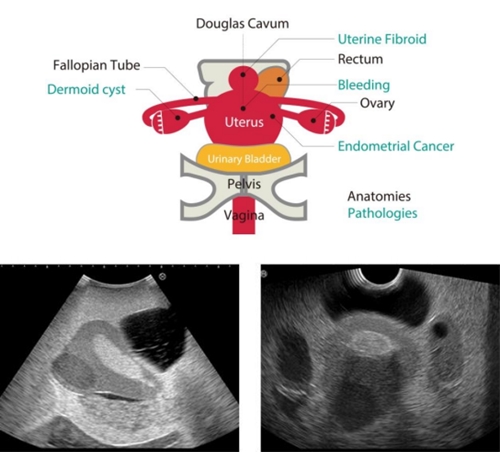
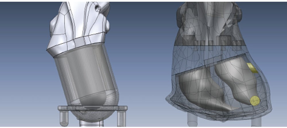
Endometrial cancer, uterine fibrosis, ovarian dermoid cyst, hemorrhage in the Pouch of Douglas.

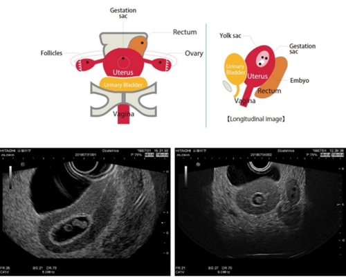
Normal pregnancy insert (7 weeks).

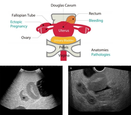
Ectopic pregnancy insert.
Ectopic pregnancy in the Fallopian tube, with hemorrhage in the Pouch of Douglas.

Training Competencies
Use and handling of transvaginal and transabdominal transducers/probes.
Imaging interpretation.
Visualization and localization of anatomy and different pathologies.
Training model for abdominal ultrasound “ABDFAN”
High-quality abdominal phantom for clinical ultrasound training, with detailed anatomy and realistic pathology simulation.
- Compatible with any ultrasound system using a convex probe.
- Accurate representation of hepatobiliary, pancreatic, renal, splenic, and vascular anatomy.
- Training capability across all eight Couinaud liver segments.
- Realistic lesion simulation: cysts, tumors (hypo/hyperechoic), gallstones, and invasive pancreatic tumor.
- Life-size and durable materials, designed for intensive training use.
Ideal for developing clinical skills from initial teaching to advanced abdominal pathology training.
Training model for breast ultrasound “BREAST FAN”
Anatomical phantom for breast ultrasound training, ideal for basic training and identification of normal and abnormal structures.
- Realistic simulation of breast tissue, lactiferous ducts, ligaments, ribs, clavicle, pectoral muscle, and lung.
- Includes targets with different echogenicities, allowing training in lesion detection and characterization.
- Visible axillary lymph nodes for practice in regional assessment.
- High-quality materials and life-size dimensions (26×38×11 cm; weight ~7 kg).
An essential tool for technical and clinical training in breast ultrasound.
SPACEFAN-ST fetal ultrasound phantom
Advanced fetal phantom for training in second-trimester ultrasound screening. Includes a 23-week fetus with realistic anatomy and a complete skeleton.
- Enables training in fetal biometry (BPD, AC, FL), anatomical assessment, and placental localization.
- Simulator uterus with amniotic fluid, placenta, and umbilical cord.
- Adjustable abdomen in four positions for versatile training.
Fetal anatomy: brain with ventricles and cerebellum, heart with 4 chambers, lungs, abdominal organs, vascular system, and external genitalia.
An essential tool for hands-on teaching in obstetrics and prenatal diagnosis.
Chest Phantom PET/SPECT
Anatomical phantom for PET and SPECT study simulation, ideal for training and verification of myocardial imaging and tumor uptake protocols.
- Myocardial uptake simulation with different radiopharmaceutical (RF) concentrations, including detection of regional defects.
- Tumor inserts in liver, lungs, and breast, with fillable spheres to test density, size, and location.
- Allows PET/SPECT image fusion with CT.

Simulated anatomy: heart (ventricles and myocardium), lungs, liver, kidneys, and breast, with specific PET “hot spots.”
A versatile tool for training, QA, and hybrid protocol development.

Anthropomorphic phantom THRA-1
The THRA-1 is an anthropomorphic phantom specifically designed for use in clinical dosimetry, allowing combined use with dosimeters.
This phantom is made of material equivalent to soft tissue, lung, and bone.
The size and spacing of the inserts for dosimeters, as well as the slice thickness, can be customized.
Head Phantom for CT Angiography (ACS)
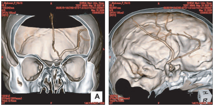
The ACS phantom was developed for the evaluation of CT angiographic images and also serves as an educational tool for medical image interpretation.

- Simulates different arterial densities according to the modality:
- Computed Tomography (CT)
- Angiography
- Metal Emission Tomography (METC)
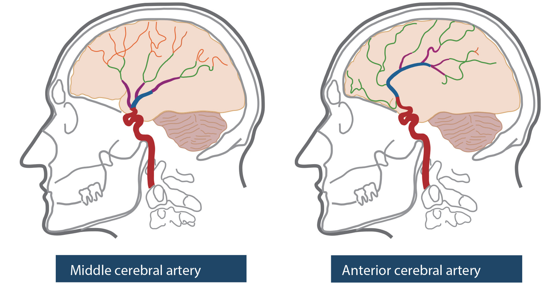
- Left cerebral arteries with integrated 3D contrast within the skull, allowing realistic and precise analyses.
Ideal for training and validation of CT angiographic techniques.
