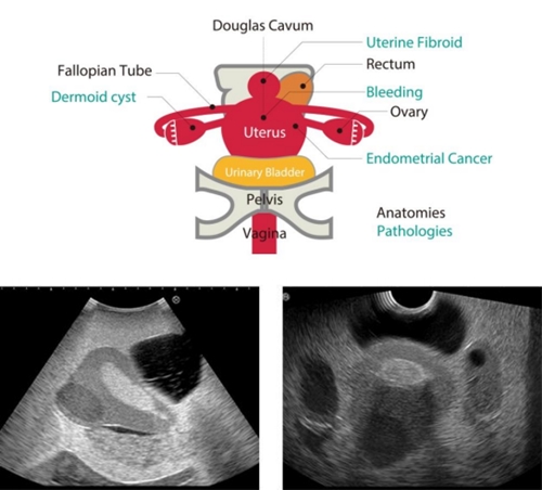
Cancro endometrial, fibrose uterina, quisto dermoide do ovário, hemorragia na Bolsa/ Saco de Douglas.
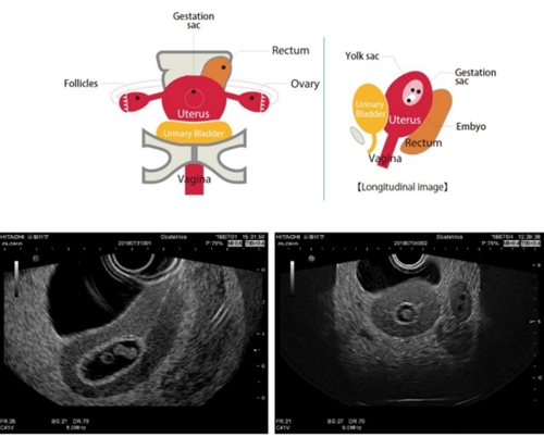
Insert de gravidez normal (7 semanas)
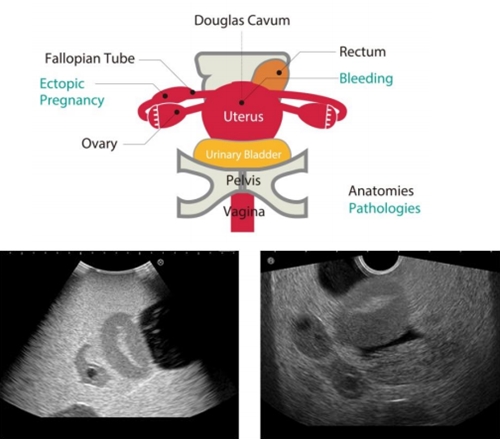
Insert de gravidez ectópica
Gravidez ectópica na trompa de Falópio, com hemorragia na Bolsa/saco de Douglas.
Competências de Formação
- Utilização e manuseamento de transdutores/sondas transvaginais e transabdominais.
- Interpretação imagiológica.
- Visualização e localização de anatomia e diferentes patologias.
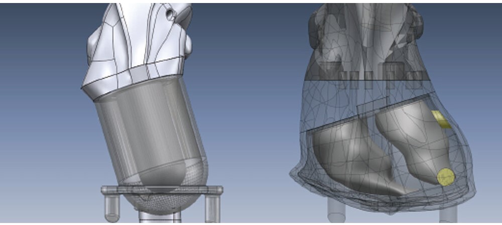
Fantoma anatómico para simulação de estudos PET e SPECT, ideal para treino e verificação de protocolos de imagem miocárdica e captação tumoral.
- Simulação de captação no miocárdio com diferentes concentrações de radiofármaco (RF), incluindo detecção de defeitos regionais.
- Inserção de tumores no fígado, pulmões e mama, com esferas preenchíveis para testar densidade, tamanho e localização.
- Permite fusão de imagens PET/SPECT com CT.

Anatomia simulada: coração (ventrículos e miocárdio), pulmões, fígado, rins e mama, com “hot spots” específicos para PET.
Ferramenta versátil para formação, QA e desenvolvimento de protocolos híbridos.
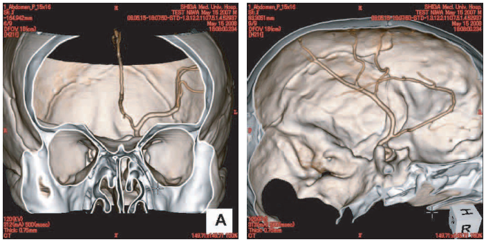
O fantoma ACS foi desenvolvido para avaliação de imagens angiográficas por TC e serve também como ferramenta educativa para interpretação de imagens médicas.
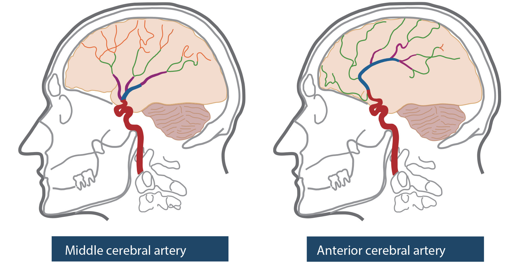
- Simula diferentes densidades arteriais conforme a modalidade:
• Tomografia Computorizada (TC)
• Angiografia
• Tomografia por Emissão de Metais (METC) - Artérias cerebrais esquerdas com contraste tridimensional integradas no crânio, permitindo análises realistas e precisas.
Ideal para formação e validação de técnicas angiográficas por TC.